PROMISE: Our kitties will never sit on top of content. Please turn off your ad blocker for our site.
puuuuuuurrrrrrrrrrrr
Jennifer Chu
Published: Wednesday, August 17, 2022 - 11:01 Ultrasound imaging is a safe and noninvasive window into the body’s workings, providing clinicians with live images of a patient’s internal organs. To capture these images, trained technicians manipulate ultrasound wands and probes to direct sound waves into the body. These waves reflect back out to produce high-resolution images of a patient’s heart, lungs, and other deep organs. Currently, ultrasound imaging requires bulky and specialized equipment available only in hospitals and doctor’s offices. But a new design by MIT engineers might make the technology as wearable and accessible as Band-Aids. 
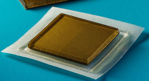
In a paper that appeared recently in Science, the engineers present the design for a new ultrasound sticker—a stamp-sized device that sticks to skin and can provide continuous ultrasound imaging of internal organs for 48 hours. The researchers applied the stickers to volunteers and showed that the devices produced live, high-resolution images of major blood vessels and deep organs such as the heart, lungs, and stomach. The stickers maintained a strong adhesion and captured changes in underlying organs as volunteers performed various activities, including sitting, standing, jogging, and biking. The current design requires connecting the stickers to instruments that translate the reflected sound waves into images. The researchers point out that even in their current form, the stickers could have immediate applications: For instance, the devices could be applied to patients in the hospital, similar to heart-monitoring EKG stickers, and could continuously image internal organs without requiring a technician to hold a probe in place for long periods of time. If the devices can be made to operate wirelessly—a goal the team is currently working toward—the ultrasound stickers could be made into wearable imaging products that patients could take home from a doctor’s office or even buy at a pharmacy. “We envision a few patches adhered to different locations on the body, and the patches would communicate with your cellphone, where AI algorithms would analyze the images on demand,” says the study’s senior author, Xuanhe Zhao, professor of mechanical engineering and civil and environmental engineering at MIT. “We believe we’ve opened a new era of wearable imaging: With a few patches on your body, you could see your internal organs.” The study also includes lead authors Chonghe Wang and Xiaoyu Chen, and co-authors Liu Wang, Mitsutoshi Makihata, and Tao Zhao at MIT, along with Hsiao-Chuan Liu of the Mayo Clinic in Rochester, Minnesota. To image with ultrasound, a technician first applies a liquid gel, which transmits ultrasound waves, to a patient’s skin. A probe, or transducer, is then pressed against the gel, sending sound waves into the body that echo off internal structures and back to the probe, where the echoed signals are translated into visual images. For patients who require long periods of imaging, some hospitals offer probes affixed to robotic arms that can hold a transducer in place without tiring. But the liquid ultrasound gel flows away and dries out over time, interrupting long-term imaging. In recent years, researchers have explored designs for stretchable ultrasound probes that would provide portable, low-profile imaging of internal organs. These designs featured a flexible array of tiny ultrasound transducers, the idea being that they would stretch and conform with a patient’s body. But these experimental designs have produced low-resolution images, in part due to their stretch: In moving with the body, transducers shift location relative to each other, distorting the resulting image. “Wearable ultrasound imaging tools would have huge potential in the future of clinical diagnosis. However, the resolution and imaging duration of existing ultrasound patches is relatively low, and they cannot image deep organs,” says Wang. The MIT team’s new ultrasound sticker produces higher-resolution images over a longer duration by pairing a stretchy adhesive layer with a rigid array of transducers. “This combination enables the device to conform to the skin while maintaining the relative location of transducers to generate clearer and more precise images,” Wang says. The device’s adhesive layer is made from two thin layers of elastomer that encapsulate a middle layer of solid hydrogel, a mostly water-based material that easily transmits sound waves. Unlike traditional ultrasound gels, the MIT team’s hydrogel is elastic and stretchy. “The elastomer prevents dehydration of hydrogel,” says Chen. “Only when hydrogel is highly hydrated can acoustic waves penetrate effectively and give high-resolution imaging of internal organs.” The bottom elastomer layer is designed to stick to skin, while the top layer adheres to a rigid array of transducers that the team also designed and fabricated. The entire ultrasound sticker measures about 2 square cm across, and 3 mm thick—about the area of a postage stamp. The researchers ran the ultrasound sticker through a battery of tests with healthy volunteers, who wore the stickers on various parts of their bodies, including the neck, chest, abdomen, and arms. The stickers stayed attached to their skin and produced clear images of underlying structures for up to 48 hours. During this time, volunteers performed a variety of activities in the lab, from sitting and standing to jogging, biking, and lifting weights. From the stickers’ images, the team was able to observe the changing diameter of major blood vessels when seated vs. standing. The stickers also captured details of deep organs, such as how the heart changes shape as it exerts during exercise. The researchers were also able to watch the stomach distend, then shrink back as volunteers drank then later passed juice out of their system. And as some volunteers lifted weights, the team could detect bright patterns in underlying muscles, signaling temporary microdamage. “With imaging, we might be able to capture the moment in a workout before overuse and stop before muscles become sore,” says Chen. “We don’t know when that moment might be yet, but now we can provide imaging data that experts can interpret.” The team is working to make the stickers function wirelessly. They are also developing software algorithms based on artificial intelligence that can better interpret and diagnose the stickers’ images. Then, Zhao envisions ultrasound stickers could be packaged and purchased by patients and consumers, and used not only to monitor various internal organs but also the progression of tumors, as well as the development of a fetus in the womb. “We imagine we could have a box of stickers, each designed to image a different location of the body,” Zhao says. “We believe this represents a breakthrough in wearable devices and medical imaging.” This research was funded, in part, by MIT, the Defense Advanced Research Projects Agency, the National Science Foundation, the National Institutes of Health, and the U.S. Army Research Office through the Institute for Soldier Nanotechnologies at MIT. First published July 28, 2022 on MIT News. Quality Digest does not charge readers for its content. We believe that industry news is important for you to do your job, and Quality Digest supports businesses of all types. However, someone has to pay for this content. And that’s where advertising comes in. Most people consider ads a nuisance, but they do serve a useful function besides allowing media companies to stay afloat. They keep you aware of new products and services relevant to your industry. All ads in Quality Digest apply directly to products and services that most of our readers need. You won’t see automobile or health supplement ads. So please consider turning off your ad blocker for our site. Thanks, Jennifer Chu covers mechanical engineering, mathematics, physics, aeronautics, astronautics, and earth, atmospheric, and planetary sciences as a writer for the MIT News Office.Stickers That Can See Inside the Body
Stamp-sized ultrasound adhesives produce clear images of heart, lungs, and other internal organs
A sticky issue
An inside look
Our PROMISE: Quality Digest only displays static ads that never overlay or cover up content. They never get in your way. They are there for you to read, or not.
Quality Digest Discuss
About The Author
Jennifer Chu
© 2023 Quality Digest. Copyright on content held by Quality Digest or by individual authors. Contact Quality Digest for reprint information.
“Quality Digest" is a trademark owned by Quality Circle Institute, Inc.