{domain:"www.qualitydigest.com",server:"169.47.211.87"} Skip to main content

User account menu
Main navigation
  • Topics
    • Customer Care
    • FDA Compliance
    • Healthcare
    • Innovation
    • Lean
    • Management
    • Metrology
    • Operations
    • Risk Management
    • Six Sigma
    • Standards
    • Statistics
    • Supply Chain
    • Sustainability
    • Training
  • Videos/Webinars
    • All videos
    • Product Demos
    • Webinars
  • Advertise
    • Advertise
    • Submit B2B Press Release
    • Write for us
  • Metrology Hub
  • Training
  • Subscribe
  • Log in
Mobile Menu
  • Home
  • Topics
    • 3D Metrology-CMSC
    • Customer Care
    • FDA Compliance
    • Healthcare
    • Innovation
    • Lean
    • Management
    • Metrology
    • Operations
    • Risk Management
    • Six Sigma
    • Standards
    • Statistics
    • Supply Chain
    • Sustainability
    • Training
  • Login / Subscribe
  • More...
    • All Features
    • All News
    • All Videos
    • Contact
    • Training

High-Resolution Method for Computed Nano-Tomography Developed

Advanced imaging technique may also be transferred to novel, laser-based X-ray sources

Fri, 10/22/2010 - 10:14
  • Comment
  • RSS

Social Sharing block

  • Print
Body

(Paul Scherrer Institute: Villigen, Switzerland) -- A novel nano-tomography method developed by a team of researchers from the Technische Universität München (TUM), the Paul Scherrer Institute (PSI), and the ETH Zurich opens the door to computed tomography (CT) examinations of minute structures at nanometer resolutions. The new method makes possible, for example, 3-D internal imaging of fragile bone structures. The first nano-CT images generated with this procedure was published in the journal Nature on Sept. 23. This new technique will facilitate advances in life sciences and materials sciences.

ADVERTISEMENT

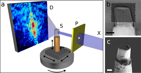
Schematic of the new nano-computed tomography (CT) method: The sample is scanned with an X-ray beam while the detector records a diffraction pattern for every beam position. The sample is then turned around its axis and scanned again, until a complete set of data is gathered for every angle. A high-resolution 3-D image of the sample is then computed from the hundreds of thousands of diffraction patterns by means of specially developed image reconstruction algorithms.

 …

Want to continue?
Log in or create a FREE account.
Enter your username or email address
Enter the password that accompanies your username.
By logging in you agree to receive communication from Quality Digest. Privacy Policy.
Create a FREE account
Forgot My Password

Add new comment

Image CAPTCHA
Enter the characters shown in the image.
Please login to comment.
      

© 2025 Quality Digest. Copyright on content held by Quality Digest or by individual authors. Contact Quality Digest for reprint information.
“Quality Digest" is a trademark owned by Quality Circle Institute Inc.

footer
  • Home
  • Print QD: 1995-2008
  • Print QD: 2008-2009
  • Videos
  • Privacy Policy
  • Write for us
footer second menu
  • Subscribe to Quality Digest
  • About Us
  • Contact Us