{domain:"www.qualitydigest.com",server:"169.47.211.87"} Skip to main content

        
User account menu
Main navigation
  • Topics
    • Customer Care
    • Regulated Industries
    • Research & Tech
    • Quality Improvement Tools
    • People Management
    • Metrology
    • Manufacturing
    • Roadshow
    • QMS & Standards
    • Statistical Methods
    • Resource Management
  • Videos/Webinars
    • All videos
    • Product Demos
    • Webinars
  • Advertise
    • Advertise
    • Submit B2B Press Release
    • Write for us
  • Metrology Hub
  • Training
  • Subscribe
  • Log in
Mobile Menu
  • Home
  • Topics
    • Customer Care
    • Regulated Industries
    • Research & Tech
    • Quality Improvement Tools
    • People Management
    • Metrology
    • Manufacturing
    • Roadshow
    • QMS & Standards
    • Statistical Methods
    • Supply Chain
    • Resource Management
  • Login / Subscribe
  • More...
    • All Features
    • All News
    • All Videos
    • Training

Self-Organizing ‘Pencil Beam’ Laser Could Help Scientists Design Brain-Targeted Therapies

MIT researchers leveraged a surprise discovery to devise a faster and more precise biomedical imaging technique

Image courtesy of the researchers

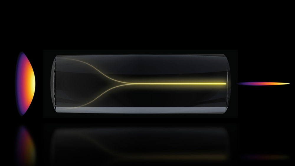
Under the right conditions, a chaotic mess of laser light can spontaneously self-organize into a highly focused “pencil beam.” This schematic shows the pencil-beam formation mechanism. Image courtesy of the researchers


Adam Zewe
Bio
Wed, 05/06/2026 - 12:02
  • Comment
  • RSS

Social Sharing block

  • Print
Body

(MIT: Cambridge, MA) -- MIT researchers discovered a paradoxical phenomenon in optical physics that could enable a new bioimaging method that’s faster and higher-resolution than existing technology.

ADVERTISEMENT

They discovered that, under the right conditions, a chaotic mess of laser light can spontaneously self-organize into a highly focused “pencil beam.”

Using this self-organized pencil beam, the researchers captured 3D images of the human blood-brain barrier 25 times faster than the gold-standard method while maintaining comparable resolution.

By showing individual cells absorbing drugs in real time, this technology could help scientists test whether new drugs for neurodegenerative disease like Alzheimer’s or ALS reach their targets in the brain with greater speed and resolution.

Concentrations of red dye begin to appear across the network veins.

 …

Want to continue?
Log in or create a FREE account.
Enter your username or email address
Enter the password that accompanies your username.
By logging in you agree to receive communication from Quality Digest. Privacy Policy.
Create a FREE account
Forgot My Password

Add new comment

Image CAPTCHA
Enter the characters shown in the image.
Please login to comment.

© 2026 Quality Digest. Copyright on content held by Quality Digest or by individual authors. Contact Quality Digest for reprint information.
“Quality Digest" is a trademark owned by Quality Circle Institute Inc.

footer
  • Home
  • Print QD: 1995-2008
  • Print QD: 2008-2009
  • Videos
  • Privacy Policy
  • Write for us
footer second menu
  • Subscribe to Quality Digest
  • About Us