Courtesy of NUS
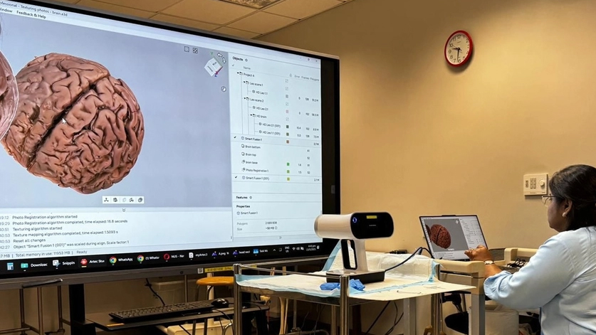
Artec Leo and 3D anatomical model of a brain in Artec Studio
I n the medical field, 3D technology is fast becoming indispensable. Whether used for educating patients ahead of surgeries or teaching students how to do these operations, anatomy models, both 3D printed and digital, offer an ideal solution for visualization ahead of time.
|
ADVERTISEMENT |
One of the main challenges behind digitizing human anatomy is accuracy. Our insides are full of complex systems and microscopic details that are difficult to capture. Blood vessels, for example, form in narrow, intricate networks vulnerable to motion artifacts during digitization.
Then there’s the question of technology integration. In theory, anatomy modeling sounds like a great interactive way of educating patients and students. But going from a captured model to usable 3D print, VR, or AR data can be tricky. File size is another issue: 3D scanning is an excellent tool for intricate object capture, but detailed models can be too heavy for VR to run.
To get around these problems, the National University of Singapore (NUS) has begun creating 3D teaching materials with Artec Leo. The wireless, target-free scanner captures human bodies in high resolution, in their entirety, in seconds, with no contact whatsoever. This makes it perfect for turning plastinated (preserved) and delicate “wet” specimens into realistic 3D models.
Ultrafast, flexible anatomy capture
Soon after Artec Ambassador Shonan 3D had set them up with Leo, NUS staff Dr. Chandrika Muthukrishnan and Dr. Arthur Lau Chin Haeng realized they’d made a wise choice. Initial skull model scans picked up everything from neck muscles to brain tissue.

NUS academic staff capturing a bone sample with Artec Leo. Image courtesy of NUS
Compared to an Artec Space Spider the team had previously used as part of a cross-university collaboration, Leo proved far more maneuverable. The cable-free device features a built-in display, battery, and processor, meaning no laptop is required during operation. Leo also gives real-time feedback, ensuring ease of operation and that no detail goes uncaptured.
Of course, the Spider boasts even higher resolution, so it’d be more than up to the task. But in this case, Leo’s versatility is vital. To capture specimens from all angles, NUS lecturers tend to suspend them from hooks in a process that would be challenging with cables in the way. Leo doesn’t sacrifice accuracy, either. The team has already achieved some outstanding results.
Dr. Muthukrishnan says, “We captured a 3D pelvic model, and our clinical consultants were very impressed with how realistic and useful it could be for explaining the details to students. The pelvic region, the anterior part of the thigh, and external genitalia are all visible—and the pelvis area, bladder, and rectum are all very important for obstetrics and gynecology.”
Getting to grips with Artec Studio
As relative newcomers, the NUS team took a little longer to master Artec Studio. But with a bit of help from Shonan 3D, they’ve learned to use its tool set for polished results. Mesh-editing essentials enable users to reduce noise, remove outliers, and fill gaps for watertight, beautifully textured models. With built-in measurement tools, it’s even possible to compare and analyze.

NUS students studying the human body using anatomical models. Image courtesy of NUS
Importantly, for VR applications, Artec Studio allows for scan decimation. This makes it possible to reduce the overall file size of captured models without losing vital details. The software also converts to OBJ and STL files at the click of a button, streamlining export for visualization and 3D printing.
According to Shonan 3D’s managing director Patrick Sng, the application potential is huge. “When you generate a 3D rendering, students are able to view it straight away from their laptops—this way of teaching has incredible potential,” Sng says. “Because of the graphics, color, and texture representation, it’s all very clear, and students get a much better understanding than they would with a physical sample.”
With access to such learning assets, students are able to manipulate real anatomical data in a 3D space, zooming in to study areas of interest, either on PC or in VR. At NUS, this approach isn’t limited to medical, either; pharmacy, life sciences, and dentistry are all areas of opportunity.
Opportunities for fine-tuning and 3D printing
NUS academic staff continues to hone 3D scanning skills. “Wet” anatomical parts stored in liquid solutions, for instance, can still be difficult to suspend. Artec Studio’s advanced alignment tools can help here, and there are many more challenging samples being lined up for digitization.

A pelvic model re-created using an Artec Leo 3D scan. Image courtesy of NUS
In the future, Dr. Muthukrishnan plans to expand this 3D modeling initiative to include head and neck anatomy as well as other complex anatomical structures, enabling medical students to better understand their intricate relationships and developmental significance. This will see the creation of more digital twins for assessment, teaching, and physical reproduction.
From there, Dr. Haeng can see VR becoming an important tool for teaching staff. “We have a bunch of VR headsets, I have an Apple Vision Pro myself,” he says. “There’s actually a software that can be connected to your mobile phone. You’re able to input 3D-scanned objects. So, these models are not just for 3D printing but VR teaching as well. We can also screencast to laptops. This would be useful in a lecture hall, for example.”
Clearly, medical study is progressing in leaps and bounds. Advanced technologies are becoming part of the everyday teaching framework. Learning experiences are getting more in-depth and interactive in a way that improves understanding and retention. As in many fields, the future is digital. At NUS, it seems that the future is now.
Published Dec. 11, 2025, by Artec 3D.

Add new comment