Featured Product
This Week in Quality Digest Live
FDA Compliance Features
Matthew M. Lowe
Don’t wait until something bad happens to make a change
Kari Miller
A lack of integration hampers quality compliance across a product’s life cycle
Etienne Nichols
A well-defined CAPA program is a framework for quality and effectiveness
Grant Ramaley
IAF CertSearch now mandatory for accredited certification bodies
Etienne Nichols
What’s the difference?

More Features

FDA Compliance News
Creates one of the most comprehensive regulatory SaaS platforms for the industry
Company’s first funding round will be used to accelerate product development for its QMS and MES SaaS offerings
Showcasing tech, solutions, and services at Gulfood Manufacturing 2022
Easy, reliable leak testing with methylene blue
Now is not the time to skip critical factory audits and supply chain assessments
Google Docs collaboration, more efficient management of quality deviations
Delivers time, cost, and efficiency savings while streamlining compliance activity
First trial module of learning tool focuses on ISO 9001 and is available now
Free education source for global medical device community

More News

Quality Digest

FDA Compliance

Opening a Cellular Door

Ultrasound delivers molecules into living cells

Published: Tuesday, April 24, 2007 - 22:00

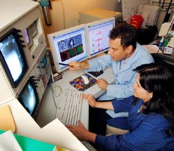
Researchers Mark Prausnitz and Robyn Schlicher use a confocal microscope to study cells whose membranes have been opened by the application of ultrasound.

Georgia Tech Photo: Gary Meek


Researchers have shown how ultrasound energy can briefly open a door in the protective outer membranes of living cells to allow entry of drugs and other therapeutic molecules and how the cells can quickly close the door themselves. Understanding this mechanism could advance the use of ultrasound for delivering gene therapies, targeting chemotherapy and administering large-molecule drugs that cannot readily move through cell membranes.

Using five different microscopy techniques, the researchers showed that the violent collapse of bubbles, an effect caused by the ultrasound, creates enough force to open holes in the membranes of cells suspended in a liquid medium. The holes, which are closed by the cells in a matter of minutes, allow the entry of therapeutic molecules as large as 50 nm in diameter, larger than most proteins and similar in size to the DNA used for gene therapy.

“The holes are made by mechanical interaction with the collapsing bubbles,” says Mark Prausnitz, an associate professor in the School of Chemical and Biomolecular Engineering at the Georgia Institute of Technology. “The bubbles oscillate in the ultrasound field and collapse, causing a shock wave to be released. Fluid movement associated with the resulting shock wave opens holes in the cell membranes, which allow molecules from the outside to enter. The cells then respond to the creation of the holes by mobilizing intracellular vesicles to patch the holes within minutes.”

Done by scientists at Georgia Tech and Emory University in Atlanta, the research was reported in the journal Ultrasound in Medicine and Biology. The work was supported by the National Institutes of Health and the National Science Foundation.

Ultrasound is the same type of energy already widely used for diagnostic imaging. Drug delivery employs higher power levels and different frequencies, and bubbles may be introduced to enhance the effect.

Scanning electron micrograph showing a prostate cancer cell immediately after exposure to ultrasound. Image has been color enhanced to show to the spot where the cell membrane has been removed.

Image courtesy of Robyn Schlicher, Robert Apkarian and Mark Baran

Transmission electron micrograph showing a prostate cancer cell immediately after exposure to ultrasound. Image has been color enhanced to show to the spot where the cell membrane has been removed.

Image courtesy of Robyn Schlicher, Robert Apkarian and Mark Baran

Ultrasound drug delivery could be particularly attractive for gene therapy, which has successfully used viruses to insert genetic material into cells, but with side effects. It could also be used for more targeted delivery of chemotherapy agents.

“One of the great benefits of ultrasound is that it is noninvasive,” Prausnitz says. “You could give a chemotherapeutic drug locally or throughout the body, then focus the ultrasound only on areas where tumors exist. That would increase the cell permeability and drug uptake only in the targeted cells and avoid affecting healthy cells elsewhere.”

Researchers have only recently found that the application of ultrasound can help move drugs into cells by increasing the permeability of cell membranes. It had been hypothesized, but not definitively shown, that ultrasound increased the permeability by opening holes in cell membranes.

Prausnitz and collaborators Robyn Schlicher, Harish Radhakrisha, Timothy Tolentino and Vladimir Zarnitsyn of Georgia Tech and Robert Apkarian of Emory University set out to study the phenomenon in detail using a line of prostate cancer cells. They used scanning and transmission electron microscopy of fixed cells and two types of optical microscopy of living cells to assess ultrasound effects and cell responses.

Beyond demonstrating that ultrasound punched holes in cell membranes, the researchers also studied the mechanism by which cells repair the holes. After the ultrasound exposure, they introduced into the cell medium a chemical not normally taken up by the cells. By varying when the chemical was introduced, they were able to determine that most of the cells had repaired their membranes within minutes.

Though the researchers used prostate cancer cells in the study reported in the journal, they have also studied other types of cells and believe ultrasound offers a general way to briefly create openings in many classes of cells.

Researchers face a number of challenges, including FDA approval, before ultrasound can be used to deliver drugs in humans. For example, the effects of the ultrasound weren’t consistent across the entire volume of cells, with only about a third affected. Researchers will also have to address safety concerns and optimize the creation of collapsing bubbles—a phenomenon known as cavitation—within bodily tissues.

Brightfield (above) and confocal/fluorescence (below) microscopy of prostate cancer cells before (left), immediately after (center) and long after (right) exposure to ultrasound. Holes in cell membranes are highlighted with an arrow.

Image courtesy of Robyn Schlicher


Before we can use ultrasound for therapy in the body, we will have to learn how to control the exposure,” Prausnitz notes. “If we can properly design the impact that ultrasound makes on a cell, we can generate an impact that the cell can deal with. We want just enough impact to allow transport into the cell, but not so much of an impact that the cell would be stressed beyond its ability to repair the injury.”

Researchers don’t yet know if the membrane holes cause long-term harm to the affected cells. General assays show that cells survive after resealing the membrane holes, but detailed studies of cell behavior are still needed. Evidence from other researchers suggests that cell membranes are frequently damaged and repaired inside the body without long-term ill effects. Cells may similarly tolerate ultrasound’s effects.

“One of the real challenges is going to be translating the successes that have occurred in the laboratory and in small animals into clinical success in people,” says Prausnitz. “Now that we better understand the mechanism of ultrasound’s effects, we can more effectively take advantage of it for medical therapy.”

This article was originally published in Georgia Tech Research News.

Discuss

About The Author

Quality Digest’s picture

Quality Digest

For 40 years Quality Digest has been the go-to source for all things quality. Our newsletter, Quality Digest, shares expert commentary and relevant industry resources to assist our readers in their quest for continuous improvement. Our website includes every column and article from the newsletter since May 2009 as well as back issues of Quality Digest magazine to August 1995. We are committed to promoting a view wherein quality is not a niche, but an integral part of every phase of manufacturing and services.