Featured Product
This Week in Quality Digest Live
FDA Compliance Features
Matthew M. Lowe
Don’t wait until something bad happens to make a change
Kari Miller
A lack of integration hampers quality compliance across a product’s life cycle
Etienne Nichols
A well-defined CAPA program is a framework for quality and effectiveness
Grant Ramaley
IAF CertSearch now mandatory for accredited certification bodies
Etienne Nichols
What’s the difference?

More Features

FDA Compliance News
Creates one of the most comprehensive regulatory SaaS platforms for the industry
Company’s first funding round will be used to accelerate product development for its QMS and MES SaaS offerings
Showcasing tech, solutions, and services at Gulfood Manufacturing 2022
Easy, reliable leak testing with methylene blue
Now is not the time to skip critical factory audits and supply chain assessments
Google Docs collaboration, more efficient management of quality deviations
Delivers time, cost, and efficiency savings while streamlining compliance activity
First trial module of learning tool focuses on ISO 9001 and is available now
Free education source for global medical device community

More News

Quality Digest

FDA Compliance

High-Resolution Imaging

Cartilage monitoring for osteoarthritis

Published: Tuesday, June 26, 2007 - 22:00

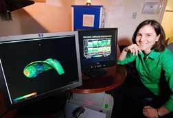
Georgia Tech student Ashley Palmer, Ph.D., conducted experiments to validate a new cartilage-imaging technique developed by associate professors Marc Levenston and Robert Guldberg in the Georgia Tech School of Mechanical Engineering. On the computer screen in the foreground is a thickness “map” of cartilage on a rabbit thigh bone generated using software associated with the new imaging technique called EPIC-microCT. On the other screen is an image showing results of EPIC-microCT scans on bovine cartilage samples at various stages of degradation.

Georgia Tech Photo: Gary Meek


An innovative combination of existing technologies shows promise for noninvasive, high-resolution imaging of cartilage in research on the progression and treatment of the common degenerative disease osteoarthritis. Microcomputed tomography (microCT), which yields three-dimensional X-ray images with a resolution 100 times higher than clinical CT scans, is commonly used to image bone for osteoporosis research, but hasn’t been useful for imaging soft biological tissues such as cartilage. These tissues simply don’t interfere with the microCT’s X-rays as they pass through a sample, and therefore they don’t show up on scans.

However, by combining microCT with an X-ray-absorbing contrast agent that has a negative charge, researchers at the Georgia Institute of Technology were able to image the distribution of negatively charged molecules called proteoglycans (PGs). These molecules are critical to the proper functioning of cartilage.

“By detecting PG content and distribution, the technique reveals information about both the thickness and composition of the cartilage—important factors for monitoring the progression and treatment of osteoarthritis,” says associate professor Marc Levenston of Georgia Tech’s George W. Woodruff School of Mechanical Engineering.

He and associate professor Robert Guldberg, also in the School of Mechanical Engineering, collaborated to establish and validate the principle of the technique, dubbed equilibrium partitioning of an ionic contrast agent-microCT, or EPIC-microCT. Then they applied the technique in vitro to monitor the degradation of bovine cartilage cores and to visualize the thin layer of cartilage in an intact rabbit knee.

“This technique will allow pharmaceutical researchers to obtain more detailed information about the effects of new drugs and other treatment strategies for treating osteoarthritis,” Levenston says.

A report on the research was published in 2006 in the journal Proceedings of the National Academy of Sciences. The National Science Foundation, National Institute of Arthritis and Musculoskeletal and Skin Disorders, and the Arthritis Foundation funded the work.

Experiments conducted by Ph.D. student Ashley Palmer established the principles and protocol of EPIC-microCT. Researchers first immersed cartilage samples in the contrast agent solution and waited for the agent to diffuse into the tissue. Tissue with fewer negatively charged PGs absorbed more of the negatively charged contrast agent, and tissue with a higher PG concentration repelled it.

Researchers then used EPIC-microCT to detect the concentrations of the contrast agent, which allowed them to calculate the amount of PGs in different parts of the cartilage. Because degrading cartilage loses PGs over time, researchers could monitor the progression of tissue changes. In addition, differences in the X-ray signal of cartilage and bone allowed researchers to isolate the cartilage layer on a rabbit joint and determine its thickness, indicating that this technique also can be used to measure tissue thinning during disease progression.

In follow-up research funded by a new, two-year grant from the National Institutes of Health, the researchers will gather additional quantitative data and use the technique to examine the very thin cartilage of rat knee joints. Researchers will nondestructively evaluate osteoarthritis progression and then attempt to use this approach to monitor cartilage changes over time in vivo inside the same live animals.

“Ultimately, if we can monitor cartilage changes with good resolution and do it with little or no invasion of the tissue in live animals, then we can track osteoarthritis progression and the effects of drug therapy or other treatments over time,” Guldberg says.

A new cartilage-imaging technique called EPIC-microCT illustrates a progressive loss of negatively charged molecules called proteoglycans (PGs) in images of degrading bovine cartilage cores (left to right, increasing degradation) and provides a quantitative measure of corresponding increases in X-ray attenuation (far right).

Image courtesy of Ashley Palmer

Researchers have already addressed a significant technical hurdle in making the imaging technique feasible. They researched several contrast agents and tried two others before choosing Hexabrix, which is approved by the Food and Drug Administration for use as a contrast agent for various imaging procedures in humans. When diluted, it produced an X-ray signal that allowed distinction of bone from cartilage.

“The ability to separate bone from cartilage in the microCT scan is a big deal,” Guldberg says. “It suggests that this technique may work in vivo.”

A new cartilage-imaging technique called EPIC-microCT yields pictures of (A) an intact rabbit knee; (B) a segmented rabbit knee with cartilage (gold) isolated from bone (red); and (C) a topographical map illustrating the range of cartilage-layer thickness in the rabbit knee.

Image courtesy of Ashley Palmer


Dilution reduces the contrast agent’s sensitivity and therefore the technique’s PG-monitoring capability, the authors write in their paper. “In this next phase of research, we hope to find a one-shot concentration of the contrast agent that works for analyzing both cartilage thickness and composition,” says Levenston, the senior author on the paper.

In addition, the researchers must address technical issues involving the in vivo delivery and retention of a sufficient volume and concentration of the contrast agent, they note in the paper.

“But even if the technique only works for in vitro studies, it still provides useful quantitative, high-resolution, 3-D images that researchers can use to nondestructively monitor cartilage degeneration and even regeneration in small animal models,” Guldberg says.

This article was originally published in Georgia Tech Research News.

Discuss

About The Author

Quality Digest’s picture

Quality Digest

For 40 years Quality Digest has been the go-to source for all things quality. Our newsletter, Quality Digest, shares expert commentary and relevant industry resources to assist our readers in their quest for continuous improvement. Our website includes every column and article from the newsletter since May 2009 as well as back issues of Quality Digest magazine to August 1995. We are committed to promoting a view wherein quality is not a niche, but an integral part of every phase of manufacturing and services.