Since the early 1900s, people in science and industry have been using a variety of optical measuring and
inspection techniques to get an enhanced view of their products for research and development (R&D). The techniques, which enable users to observe, measure and analyze parts for quality
control purposes, have moved out of their original homes in manufacturing environments and into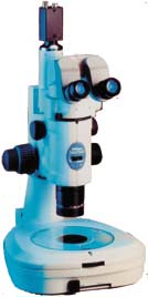 the laboratory. the laboratory. This article is intended as a simple introduction to optical measurement. Readers familiar with the material
contained in sections of this overview will find it a useful review of the basic principles of the field. Others who are using or purchasing optical measuring equipment for the
first time may find it helpful in explaining the issues to consider in selecting and purchasing the appropriate instrumentation to meet their needs. Measuring and inspection R&D facilities usually have to employ both inspection
and measurement in evaluating samples. The two evaluative methods give quite different information. Inspection is designed to answer qualitative questions,
such as "Is this flat enough?" or "Are there too many pits in this tool?" or "Do the grain and color of this sample
look right?" or "Is this contaminated?" Measurement is designed to answer quantitative questions, such as "How long is this piece?" or
"What is the thread pitch of this screw?" or "How deep is this hole?" or "How far apart are the centers of these grooves?" Some questions probe for information that
can be considered semi-quantitative, such as "Does this match my overlay?" or "Does this line up at the right place?"
Many R&D environments use measurement tools to get quantitative information for developing engineering prototypes or final production-line products or evaluating
defects in existing materials or products. In life-science R&D labs, measurement can validate accuracies of diagnostic products or the usability of prosthetic devices.
Optical measuring uses human eyes to compare specific sample features against standards that are set in advance. It's often just one of many evaluative techniques
in use in the facility, including various test, inspection and calibration techniques. But it's especially significant because in most R&D environments there's no
substitute for visual information in ascertaining future performance potentials for a product or material.
Selecting the right measuring techniques Selecting the right technique for measuring in a given environment is an important
decision that must reflect the user's specific needs. Depending on the sample size, necessity for color or 3-D accuracy, the need for computer interface with designers
or others on the development team, specific parts to be measured, tolerances allowed, and throughput required, R&D labs will select appropriate techniques for
handling their optical measuring needs. In many R&D laboratory situations, microscopes are selected because at low throughput levels they offer the highest
accuracy and best color fidelity of any method of optical measuring. Other important technologies include optical comparators and video-based measuring systems. What is a microscope? In its most basic form, a microscope starts with two simple magnifying lenses,
placed a specific distance apart and a set distance from the eye of the observer. Light illuminates the sample, creating an image that passes through the first lens,
where it's magnified. The magnified image then passes through the second lens, which magnifies it again. Then it passes on to the user's eye. To determine how
much total magnification a system offers, multiply the magnification provided by each lens. For example, if each of the two lenses provides 10X magnification, the total magnification is 100X. In most microscopes, the primary lens, the one closest to the specimen, is called the objective. This is the lens you select from a revolving circular turret, or
nosepiece, on the instrument. The microscope also has secondary lenses, usually those closest to the eye, which are located in the eyepieces. Many microscopes
have condensers and other additional lenses that serve specific purposes. But a light source and lenses are just the beginning. After all, a magnified image
must be a true representation of the specimen in detail, shape and color. So microscopes have many other parts, all of which contribute to the quality and
fidelity of the image you see. Filters, diaphragms, lens coatings, special accessories and techniques for observation all affect the raw image so the user gets better, more accurate information.
Common microscope illumination techniques
There are many techniques for illuminating microscope samples for measurement. Most R&D applications use either or both of the following techniques:  Brightfield is an illumination technique that provides flat, even illumination of the
field of view. Switch the microscope to brightfield illumination and you'll clearly see where things are located, how they are attached to each other, and how the color
and grain of the sample appear. Brightfield illumination is used to view cracks, discoloration, contamination and dirt, as well as to monitor assembly and orientation of components. Brightfield is an illumination technique that provides flat, even illumination of the
field of view. Switch the microscope to brightfield illumination and you'll clearly see where things are located, how they are attached to each other, and how the color
and grain of the sample appear. Brightfield illumination is used to view cracks, discoloration, contamination and dirt, as well as to monitor assembly and orientation of components.
Darkfield illumination lights the specimen surface from an oblique angle, so dirt, contaminants, pits in the finish, deviations in flatness, scratches and other surface
problems are strikingly clear. Darkfield is used for observing the surface, looking for contamination and checking for deterioration in a product or part. In the R&D
environment, darkfield illumination is used to identify and locate X-Y coordinate pairs and measure the dimensions of defects found in prototypes or production models for reengineering. Other microscope illumination techniques are designed to meet specific needs in specialized R&D environments. These techniques include:
Polarized light and differential interference contrast (DIC) are techniques that use polarizing materials and/or optical prisms to examine physical and structural
characteristics of manufacturing materials. For example, they're used in examining unprocessed semiconductor wafer substrates, ceramics, crystals and metal alloys.
In biomedical labs, DIC is used to optically section individual cells and cell structures. Fluorescence is a phenomenon whereby a chemical excited at one light wavelength emits light at a different and longer (usually visible light) wavelength.
It's used in the semiconductor industry, for example, to identify and measure the extent of contamination in photoresistant chemicals. In the life sciences, new
antigens and antibodies are often identified by means of this technique. Phase contrast takes advantage of the unequal transmittance of light through a structure based on the different densities of the structure's components by using a
phase ring in the objective lens and a phase annulus in the condenser assembly of the microscope. Phase contrast is commonly used in biomedical research and
development to examine unstained materials, either living or dead. Special issues for microscopic measuring
There are two basic ways of measuring with a microscope: field-of-view measurement and stage-movement measurement. Field-of-view measurement can
use small, precise eyepiece reticles that superimpose a pattern or scale over the image. It can also provide very accurate quantitative measuring because there are no
stage-movement errors. If the entire region to be measured can be seen at one time in the eyepieces, this is a quick, precise way of measuring.
Stage-movement measurement is used when the feature to be measured doesn't entirely fit in the field of view or when a sample within the field of view must be
moved past a fixed point for measurement. Typically, a linear scale or a rotary encoder inside a drum micrometer is used to measure the displacement of the stage
as the object on the stage moves past a reference point in the eyepiece (typically a crossline).
A linear scale registers a direct measurement of how much the plates in the stage move and provides a digital readout that is extraordinarily accurate--down to less
than a half micron (0.00002 in.). In contrast, a drum micrometer measures the turning of a gear that moves a
spindle, which in turn moves the stage. The measurement is read off of a graduated drum and a vernier scale. Although these measurements are not as accurate as those
provided by a linear scale, they are more economical and are certainly quite sufficient for many users' needs.
Whatever kind of measurement you are doing, it's important to have the best optical resolution for feature detection. In addition, the stand of the microscope should be
massive and stable to eliminate vibrations that can affect the accuracy of the measurement. Confocal imaging
One very important development in measuring for the research environment is confocal microscopy. This requires a microscope different from either the standard
compound microscope or the stereomicroscope and is based on a completely different kind of optical theory. Confocal microscopes use white light or lasers to
construct a highly detailed "map" of a 3-D sample. Simply put, they optically section a given sample point-by-point and layer-by-layer. They can completely reconstruct
the object on a computer screen, and allow you to rotate the image and see your component or material from any angle.
Microscopes vs. optical comparators Microscope measuring offers the best resolution, the greatest optical versatility and
the broadest range of magnifications of any optical measuring technique. In addition, microscopes are usually assembled in a modular "building block" design,
allowing the lab extraordinary flexibility in putting together an optical system optimized for its specific requirements. Measuring microscopes offer users the
choice of episcopic (reflected), diascopic (transmitted) or oblique (off-axis) illumination, or a combination of these, depending on need. They also have
unparalleled documentation capabilities for photo and video, and most microscope systems can be upgraded for video after purchase if desired.
On the other hand, microscopes are best for "small scale" work; they offer a smaller field of view, are designed for smaller sample sizes and offer limited stage
travel. In addition, the same eyepieces that provide the optimal resolution and accuracy can lead to eye fatigue if not adequately adjusted for comfort over long periods of use.
Optical comparators, also called profile projectors, use large ground-glass screens for imaging. They offer a much larger field of view and cause less eye fatigue over
long usage. In addition, comparison overlays can be used with them to provide go/no-go information for specific samples.
The drawbacks to optical comparators is that they offer somewhat less resolution and are relatively limited in terms of the types of detailed documentation they can
provide. Optical comparators are also large--too large for some cramped lab environments. Video measuring
In some ways, video imaging offers the best of both worlds. With its excellent online documentation and archiving capability, repeatability, and capacity for image
processing and manipulation, a video-based system offers extraordinary capabilities. A fully automated video measuring system will find randomly oriented parts with a
computerized vision system, adapt to their varying orientations, take a prescribed series of measurements and compare those measurements to your preset tolerances for evaluation.
Video has other advantages as well. For instance, it can be used not only for measuring, but also for inspection, teaching and documentation. Furthermore,
because the user is looking at a monitor, these systems can be extremely comfortable to use over long periods of time. Finally, they can offer a graphical part
display--a "road map" for measuring sequences. Video-based microscope measuring systems are becoming increasingly prevalent in government and industry, and may
one day be the standard for taking measurements. Shopping for a measurement system
As is the case with most purchasing decisions, there's a relationship between price and performance in the purchase of a measuring system for the R&D environment.
An instrument can be purchased for as little as $100 or as much as $200,000, and the typical sale for a precision measuring system with a digital readout can range
from $5,000 to $50,000 or more. Video measuring systems range from $15,000 to more than $100,000 in price. Here are some questions to ask when comparing measuring systems:
Am I going to use this instrument just to measure or to inspect as well? If you need to measure small features in the X-Y plane, you should probably buy a
compound microscope. In most cases, a dedicated measuring microscope with a measuring stage is best for this purpose. Do I need to see features three-dimensionally? If you need to see very
sophisticated 3-D images at high magnifications, you'll want to look into a confocal attachment for your microscope. What requirements do I have for the field of view? If you need to measure large
components, it helps to have an instrument with a large field of view, such as an optical comparator. How big are my samples? The depth of the specimen determines the working
distance you need. The working distance is the amount of space between the surface on which you are focused (the focal plane) and the front element of the
objective lens. Generally speaking, the greater the working distance you require, the less magnification you can achieve, and the lower the overall resolution of your
system. If you have 3-D specimens, look for lenses with the highest numerical aperture (NA). High-NA lenses give the best resolution over long working distances.
Also, consider the X-Y size of your samples. When measuring, you will be limited by the distance the stage can travel in each direction. Samples that are too large for
measuring microscope stages are best suited for optical comparators. What are my current and future needs? Look for a system that is modular and adaptable. For instance, you may only need reflected light right now, but eventually
may want to add transmitted light. You may do two-axis measuring now, but eventually may want to measure in three axes. Or, you may not need to document
your findings now, but eventually may want to record your findings by taking photomicrographs or using video. Look not only at today's needs, but at tomorrow's
as well. If you buy a nonmodular system, you may save now only to find your system outdated in a few years. Is the manufacturer a widely respected company? No one company makes the
best of everything. Look for a company whose instruments have a reputation for top optical quality and can be retrofitted to meet your changing needs. Also, make
sure the dealer's salespeople have the service orientation and the field knowledge necessary to become your partners. Which system for you? Measuring microscopes, optical comparators and video-based systems can all be
effective tools for quantitative measurement in R&D applications. The essence of a measuring system is its optics, and there is never a substitute for optical integrity.
The "garbage in, garbage out" is never truer than when referring to optics-based measuring instruments with inferior image quality. Buy the best optics you can
afford. True measuring optics are highly corrected for both spherical (shape) and chromatic (color) aberrations and will introduce no errors into a quantitative measuring system.
Finally, the instrument you select today should be optimized for the tasks for which you intend to use it. An instrument that has a well-conceived design will not only
fulfill current requirements, but also can be easily upgraded as your needs become more demanding. About the authors
William Chambers is product manager specializing in optics and optical theory for industrial microscopes at Nikon Inc., He has a degree in biomedical photographic
communications from RIT. He has been with Nikon since 1988. Jack Isaacson is a Nikon field sales engineer for metrology products. An 18-year
employee of Nikon, he is an experienced trainer, presenter and educator on the subject of metrology tools and techniques. For more information on this article or
on Nikon products, contact Nikon Inc.; phone (800) 526-4566; or e-mail wchambers@qualitydigest.com . |