by Steven A. Mango
From Dinosaurs to Pipelines
Dinosaurs may pose interesting problems for radiography, but those problems aren't unique. Pipeline radiography has its own set of problems. For example, how do you use radiography to test welded seams and other pipe materials over miles of pipeline?
Praised as highly efficient X-ray tools in the field, X-ray crawlers are self-contained, self-powered exposure vehicles regularly used for internal radiography of large-diameter pipes. Crawlers may be traditional X-ray inspection equipment mounted to custom-built remote control vehicles or they may be off-the-shelf equipment designed to function in a variety of pipe diameters and environments. Functioning like a remote-control car, this distinctive apparatus fits inside pipelines and crawls along their inner surfaces, entering and exiting at specific access ports.
Highly versatile X-ray tools, crawlers work in a wide variety of conditions for nondestructive examination of pipeline welds, corrosion monitoring of pipeline walls and other pipeline integrity measurements. Many are designed to produce high-quality panoramic radiographs of circumferential welds in both onshore and offshore pipelines. More advanced crawlers work virtually independently of their operators and aren't reliant on being tethered to an operator.
For tracking purposes, technologists attach low-activity isotopes to a crawler's body to monitor location, speed and direction. This also allows the radiographer to control and manipulate crawlers on command without physically seeing them. Thus, operators can dictate how crawlers move, what they radiograph and when it all happens.
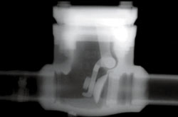
Even though X-ray crawlers are designed to cope with varying pipe diameters and wall thicknesses, certain factors can make crawlers difficult to pull in and out. Unfortunately, older pipe networks usually lack any conveniences that help X-ray inspectors do their jobs. It's occasionally necessary to remove massive valves or fittings before a crawler can be inserted into a pipe.
Crawlers are excellent tools for examining pipelines from the inside out. But that isn't always practical or needed. When it's necessary to perform X-ray inspections from the outside of a pipeline and in a confined area, gamma sources are helpful tools. These small, portable radiation sources are good for testing pipelines in refineries or power plants, where accessing inspection areas can be challenging and even dangerous.
Pipe workstations are often in motion, complicating work environments and increasing the potential for injury. For pipe networks, workers must inspect welds one by one, along the length of the pipe, using mobile X-ray units. Clearly, this is far more problematic than working at fixed locales. Mobile X-ray units fit the bill for these applications.
Although the mobile X-ray unit comes in a number of models, the configuration most widely used in pipeline and refinery testing is the tried-and-true truck-mounted unit. This setup has its advantages, but issues of durability, storage, and lighting still present troubling obstacles.
|
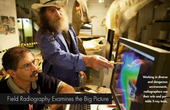
As methods in digital X-ray technology advance, nondestructive testing (NDT) applications continue to present interesting challenges to technologists in the field. When it comes to real-life practice, on-site radiographers sometimes confront problems that require resourceful solutions--especially when challenged with radiographing a 77 million-year-old mummified dinosaur in the badlands of Malta, Montana.
On July 27, 2000, in Montana's badlands, a Judith River Dinosaur Institute dig team led by famed paleontologist Nate Murphy uncovered an amazing find: a 90-percent-intact Brachylophosaurus canadensis. Estimated to have been 22-feet long and weighing about 1.5 tons when it died, Leonardo, as the duck-billed dinosaur was dubbed, was extracted intact in a 6.5-ton block after months of painstaking excavation. It is one of only four dinosaur specimens in the world with fossilized skin and muscle.
When the Judith River scientists wanted to uncover the secrets within the remarkably well-preserved mummified dinosaur, they called on Eastman Kodak Co.'s nondestructive testing division and its Michigan-based customer, NDT Group Inc.
Few dinosaur fossils discovered before Leonardo bear scales and tissue parts, but about 90 percent of Leonardo's skeleton is covered in fossilized soft tissue, including muscle, nail material and beak. Leonardo's body was so unusually mummified that even its stomach and its last meal were remarkably well preserved, earning Leonardo the distinction of "Best-Preserved Fossil in the World" from Guinness World Records.
Paleontologists wanted a closer look inside Leonardo. Ordinarily, radiographic equipment doesn't provide sufficient power and resolution to do the job, or the mobility to get to the fossil. During one intense week in late June 2006, JRDI invited a team of Kodak and NDT Group engineers to bring a complete digital radiography lab to the field station in Malta. It was there that paleontologists and radiographers employed modern science and nondestructive evaluation (NDE) methods to learn more about Leonardo.
The Kodak and NDT Group team worked with JRDI's scientists to produce more than 40 select radiographic images of Leonardo's head, portions of his skeleton, and abdomen using computed radiography (CR) techniques. The digital radiography lab included the Kodak Industrex ACR 2000i Digital System, high-powered X-ray tubes, and iridium and cobalt gamma sources.
Because CR of an entire mummified dinosaur isn't an everyday occurrence, there was some concern on the part of the scientific team about how well such a technique would work. In addition, this radiographic application was the first of its kind, so scientists had no idea what to expect.
Because electromagnetic sources vary in strength and intensity, it's important to choose the right source for the job. To begin the unprecedented examination, Kodak and the NDT Group performed radiographic testing on Leonardo with a relatively small, 160 kilovolt (kV) X-ray tube, restricting testing to fairly thin portions of the specimen.
To penetrate the denser portions of Leonardo's skin and bone, radiographers set up a larger (300 kV) X-ray tube, as well as very powerful Iridium-192 and Cobalt-60 gamma sources. Iridium-192 emits a range of gamma rays at energies from 137 kV to 651 kV, while Cobalt-60 emits gamma rays at 1.17 and 1.33 megavolts (MeV). Radiographers required these higher-energy gamma sources to penetrate very thick, compact material, such as portions of Leonardo's skull and beak.
In Leonardo's case, radiographers employed what they had on site--two X-ray tubes and two gamma sources. The team worked on various skin thicknesses of the fossil with both sources, depending on how much energy it took to penetrate the surface.
The best results were obtained using digital imaging plates and the lowest-energy (160 kV) X-ray tube to image the thinnest portions of the specimen. Although high-energy X-rays and gamma sources were needed to look at thicker sections of Leonardo, these images are grainier than those from the low-energy sources. In addition, the high-energy sources used on the thicker portions of Leonardo produced more scatter of the primary beam. Both of these factors are detrimental to image quality. Despite the tradeoffs between penetrating power vs. image quality, the CR system still pulled off some remarkable images, many showing an amazing amount of detail in the fossilized tissue.
With field NDT, you often don't know what your space constraints are going to be until you try to set up your equipment. Obstructions in the field of view, limited area for equipment and being able to set up an adequate radiation safety zone mean field technicians have to be ready for anything. One of the biggest challenges encountered at Malta was shooting around the fixed steel frame supporting Leonardo's massive body. The immense weight of the fossil kept it from being moved, and there was little room underneath it to fit an X-ray tube.
During the imaging process, team members often had to compromise the best shots and angles to avoid capturing the structure. The steel obstructions limited where workers could place their X-ray equipment, and caused excessive scatter--an unwanted signal that produces poor image quality. Because radiographic inspection wasn't considered when constructing the steel support, its sheer thickness blocked any hope of passing electromagnetic rays through the fossil. Unwilling to scale back research efforts and miss encroaching deadlines, NDT teams scurried to find a way around this particular obstacle. To limit the scatter of X-ray beams, technicians constructed lead shields and placed them close to inspection areas.
A further complication was that the negative air chamber housing Leonardo was very small and left little room for movement. Depending on the application, on-site radiographers sometimes use gamma sources to generate electromagnetic rays in tight confines. Popular for field applications, gamma sources are versatile and extremely portable--about the size of a soup can. Fortunately, the team had brought gamma sources for their penetrating power. Their compact size was an added bonus that helped researchers do their work in the confined space. To have more flexibility when arranging shots, the team rigged a boom device using a pole and spare ladder. This allowed radiographers to fix gamma sources over the dinosaur, giving them the option of shooting above the specimen with imaging plates secured underneath.
For a variety of image angles, radiographers also wanted to shoot Leonardo from below. To get around the steel frame, the team needed to cut away portions of the frame. Using a high-powered hydraulic jack, the team lifted the frame (still supporting the fossil) off the ground. Once it was firmly elevated, a welder cut away portions of the frame without weakening the support. This gave radiographers space to slide their imaging equipment underneath Leonardo, making room for some very scientifically interesting shots.
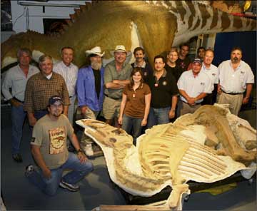 The computed radiography team with Leonardo and full-size model of Brachylophosaurus canadensis in background.
|
In field radiography, physical shielding can be hard to come by. Because radioactive materials can never be turned off, safely managing the gamma source was a constant responsibility for the Leonardo team, challenging them to think creatively about securing hazardous work areas.
For safety reasons, it was imperative to manage personnel in and around the workstation. To do this, the team lined up several rental cars from front to back, creating an impassible barricade that kept onlookers a safe distance away. To minimize emissions, NDT engineers constructed a bunker around the X-ray sources using the field station's inventory of 50-lb bags of calcium carbonate.
Both solutions helped limit human exposure to high doses of radiation; however, additional shielding was needed to prevent gamma radiation from reaching a nearby intersection. One call to the city highway department produced a quick solution; the city delivered a huge front-end loader and parked it adjacent to the building nearest to the source. That weakened the radiation enough to safeguard the intersection.
For field NDT operations, time constraints are almost always an issue. Whether shutting down a production area to perform X-ray inspections or shutting down a paleontology lab to examine a dinosaur, managing workloads and estimating timelines is always more difficult to do in the field. Very few predictions prove accurate in the field because it's hard to foresee what will come up ahead of time. For Leonardo's team, time was of the essence. X-ray technicians had only three days in which to work, and that forced one more quick solution.
As deadlines approached, the team chose to accelerate the imaging process on the last day by doubling plates--a technique that produced two side-by-side images in one exposure, cutting process time in half.
The Leonardo Project was an astounding success. Radiographers were amazed at how well the digital imaging technology performed. This gave the NDT team reason to celebrate, but even more valuable is what Leonardo has done for the scientific community. Presently, word of this one-of-a-kind application and the versatility of digital radiographic testing are starting a new era of nondestructive testing within the world of paleontology.
To learn more about the experience and imaging aspects of The Leonardo Project, read the technical paper available in the February 2007 issue of Materials Evaluation magazine. In spring 2008, The Discovery Channel will air an in-depth documentary on the discovery of Leonardo and the mysteries behind the world's best-preserved dinosaur.
A Kodak employee for more than 30 years, Steven A. Mango joined the company's nondestructive testing group in 2002. He currently serves as worldwide technical manager, overseeing all regional activities dealing with Kodak's nondestructive testing products. Mango also manages the latest technologies in computed radiography at Kodak's state-of-the-art demonstration lab located in Rochester, New York.
Mango has written, presented and published several papers on various aspects of computed radiography and is an active member of American Society of Nondestructive Testing and ASTM International. He's a graduate of Rochester Institute of Technology and earned his bachelor's degree in image science.
|