Social Sharing block
A new research center designed to examine materials and structures across many length scales has been launched at the University of Southampton in the United Kingdom. The µ-VIS (micro-vis) X-ray Imaging Center examines the internal structure of objects in incredible detail. It produces high-resolution 3-D images that support research in fields ranging from biomedical science to engineering, and archaeology to modern environmental science.
|
ADVERTISEMENT |
The £2.2 million ($3.4 million) research facilities provide micro-focus computed tomography (CT) imaging to exceptional energy and length scales (up to 450 kV, imaged volumes in excess of 1 cubic meter), while offering complementary resolution capabilities down to 200 nm.
|
|
Looking inside objects at very detailed levels
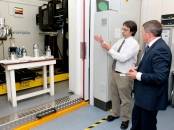
Inaugurated on September 16, 2011, Southampton’s µ-VIS Imaging Center is equipped to achieve breakthroughs in the engineering, biomedical, environmental, and archaeological sciences. The center integrates state-of-the-art imaging hardware, world-class computing, and image processing expertise to acquire and process the 3-D data that are needed to break new ground.
Three systems installed at the center are from Nikon Metrology, namely a custom-designed Hutch, with a 450 kV and 225 kV source as well as a flat-panel and line-array detector, a modified XT H 225 ST cabinet system, and a 160 kV Xi Benchtop scanner. The center additionally incorporates a Gatan XuM/Zeiss Evo MA25 nanoCT system and a SkyScan1176 for in vivo CT (e.g., small animal studies).
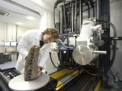
The µ-VIS scanners work in much the same way as a medical CT scanner, but at higher resolutions, by taking thousands of X-ray views to build up a 3-D image of the examined object. Beautifully rendered CT images illustrate the scanner’s capability in measuring internal and external dimensions as well as the critical insight it provides through the additional fourth dimension of material density. Looking inside objects at this level of detail in a nondestructive way is a huge advantage when studying objects that either cannot be dismantled or are too unique, delicate, or complicated to take apart.
|
|
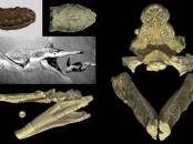
A ferocious predator’s jaw
The opening seminar at the center’s inauguration event was organized as a symposium focusing on application results. Professor Ian Sinclair, the head of the µ-VIS Center, discussed the Weymouth Pliosaur research where a 2.5-m-long jaw has been scanned and reconstructed using the custom Hutch system.
The huge Nikon Metrology X-ray bay is ideal for scanning such large, dense lumps of fossil. The X-rays are helping to build up a 3-D picture of this ferocious predator, which terrorized the oceans 150 million years ago. The pliosaur was an aquatic reptile with a huge bulky body, paddle-like limbs, and an enormous crocodile-like head packed full of razor-sharp teeth. By looking at the inner architecture of the skull, scientists may gain new insights into the species and its evolution.
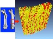
Other natural-specimen studies include the scanning of large numbers of mice as models for numerous human conditions, including osteoporosis. Researchers have, for example, recently used the XT H 225 ST equipped with a transmission target to scan the bones of gene knock-out mice, elucidating for the first time the critical role of certain proteins in bone fragility. The pore analysis method applied illustrates how much insight could be gained through creative X-ray image and CT slice post-processing.
Important issues for future farming were investigated via quantification of the structure of soils and development of living plant roots, supporting multiphysics modeling of how this may affect both irrigation and phosphate utilisation.
|
|
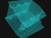
X-ray makes the difference in engineering insight
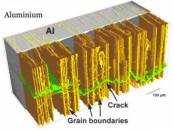
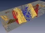
Industrial engineering benefits greatly from the broad application reach of CT scanning. Groundbreaking research involved the study of carbon fiber and epoxy used to coat aluminum gas bottles to reduce weight. The research looked into the resistance to damage, and used all scales of the µ-VIS Imaging activities. The full engineering components were initially scanned in their intact form, with subsequent targeted higher-resolution subsampling that elucidated structures at individual ply, tow, and, ultimately, discrete carbon-fiber levels. This work has allowed established theories of fiber failure modes to be explicitly compared with experimental results for the first time at both coupon and engineering component levels.
A railway engineering study reported on the effect of long-term use of the ballast underneath railway sleepers (railroad ties). The crushed-stone ballast facilitates drainage of water and distributes the load from the railway sleepers. Taking several scans of such a dense material at meaningful sample length scales (sample in the order of 300-400 mm diameter) confirmed that critical data on individual particle rotations could be obtained in acceptable time scales using the custom hutch's curved linear array and 450 kV source. The results are now being taken forward to validate discrete element method (DEM) simulations of ballast deformation.
More information and images can be found at http://www.soton.ac.uk/muvis/.
Add new comment